Hayve 1600X Digital Inspection Microscope
Official Store Deal
Expert Analysis Overview
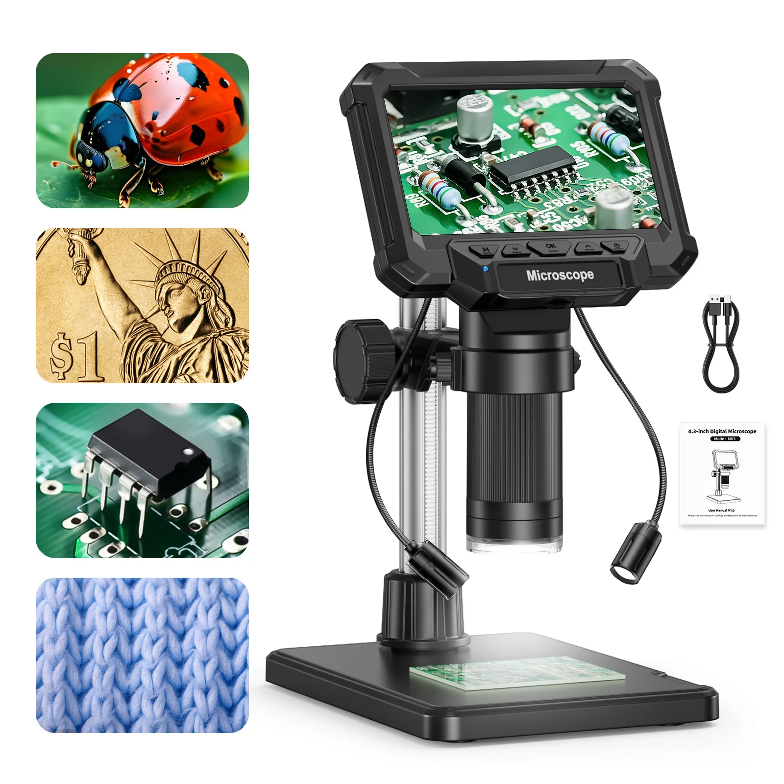
The Hayve 1600X Digital Inspection Microscope is a highly capable optical instrument designed for detailed visual analysis across a broad spectrum of micro-scale applications. This device targets hobbyists, educators, and light professional users who require precise magnification and clear digital output for tasks such as circuit board inspection, numismatic examination, and botanical observation. Its integrated display and robust feature set position it as a significant upgrade over traditional handheld magnifiers, offering enhanced functionality and improved data capture. This is a serious tool.
The core utility of any microscope lies in its magnification prowess. This Hayve unit provides a 50X to 1600X magnification range. This broad spectrum allows for a versatile approach to specimen analysis, accommodating both broad overviews and granular examinations. The range is impressive.
At the lower end, 50X magnification is ideal for general overview. It provides a wide field of view, making it simple to orient oneself on larger objects. This is useful for quickly locating areas of interest on larger objects like printed circuit boards or entire coin surfaces, where a broader context is initially required before zooming into specific points. The ability to scan a broader area efficiently saves considerable time during initial inspection phases, preventing the user from getting lost in excessive detail too soon. This initial scan is fast.
Transitioning to the higher end, the 1600X magnification reveals intricate details with remarkable clarity. Imagine examining the minute solder joints on a surface-mount device, where even a slight imperfection can lead to circuit failure. Or perhaps the subtle wear patterns and minting errors on a rare coin, which can significantly impact its value. This level of zoom brings forth features that are otherwise imperceptible to the naked eye or even lower-powered magnifiers. It allows for the identification of micro-fractures, corrosion, or even the precise alignment of individual fibers in a textile sample, crucial for material science or forensic analysis. The resolution at these higher magnifications is critical for accurate defect identification and detailed characterization. Fine details emerge.
Compared to entry-level optical microscopes, which often cap out at 400X or 1000X, the 1600X capability of this digital model offers a distinct advantage for observing finer structures without the complexities of traditional setups. Traditional optical systems attempting such high magnifications typically require oil immersion lenses, adding complexity, mess, and recurring costs due to the specialized immersion oil. This digital alternative simplifies the process considerably. It delivers comparable visual detail without the need for specialized consumables or intricate lens changes. The digital sensor captures and processes the image, providing a clear, magnified view directly on the screen, making high-power observation more accessible. It's a cleaner solution.
Central to the user experience and the immediate utility of this instrument is the 4.3-inch IPS adjustable screen. This display is not merely a passive viewing portal; it is an interactive interface that significantly enhances usability. The screen is key.
The IPS (In-Plane Switching) technology ensures wide viewing angles. This means that color accuracy and image clarity remain consistent even when viewed from oblique positions, up to 80 degrees of adjustment. This is particularly beneficial in collaborative settings, such as a classroom or a workshop, where multiple individuals might need to observe the specimen simultaneously without significant degradation in image quality. The screen's adjustability, allowing for tilt and rotation, further enhances ergonomic comfort. Prolonged inspection sessions become less fatiguing, as users can position the screen to suit their individual posture and line of sight, reducing neck and eye strain. Comfort is paramount.
A common frustration with many entry-level digital microscopes is a fixed, often low-resolution, TN panel display. This often necessitates immediate connection to an external monitor for any serious work, adding an extra layer of setup and potential compatibility issues. The integrated 4.3-inch IPS screen on the Hayve unit mitigates this significantly. It provides a sufficiently clear and vibrant image for most standalone inspection tasks, making it a truly self-contained unit. The convenience of an all-in-one unit cannot be overstated; it simplifies setup, reduces cable clutter, and allows for immediate operation out of the box. It works right away.
This integrated display offers a significant upgrade over models that rely solely on smartphone or PC connectivity for viewing. Such reliance introduces potential latency, software compatibility issues, and the need for additional devices. The dedicated screen ensures immediate visual feedback, with minimal lag, and provides a stable, consistent viewing platform. This is a critical factor for maintaining focus during delicate inspection procedures, where even a slight delay can disrupt the workflow. The display is reliable.
Effective illumination is paramount for clear microscopic observation, directly impacting the visibility of fine details and surface textures. This unit incorporates 8 built-in LED lights. These are strategically positioned around the lens to provide optimal coverage. Lighting is crucial.
The array of LEDs provides uniform, shadow-free lighting. This is crucial for revealing subtle surface textures and topographical features that might otherwise be obscured. A single, directional light source often casts harsh shadows, which can obscure important details and create misleading visual artifacts. The multi-point LED system minimizes this effect, ensuring consistent illumination across the entire field of view, regardless of the specimen's contours. The brightness is adjustable via a light wheel, allowing users to fine-tune the intensity to suit specific observation needs. Different specimens require different lighting conditions; a highly reflective metallic surface, for instance, benefits from diffused, lower intensity light to prevent glare, while a translucent biological sample might require brighter, more direct illumination to enhance internal structures. Control is precise.
Compared to microscopes relying on external, often cumbersome, light sources or fewer, less powerful LEDs, this integrated system offers superior control and consistency. The absence of external light sources reduces clutter on the workbench, creating a cleaner and more organized workspace. It also streamlines the setup process, as there are fewer components to manage. The ability to precisely control illumination directly from the device enhances the overall analytical capability, ensuring optimal contrast and clarity for diverse materials and observation objectives. This integrated approach is efficient.
Beyond live viewing, the microscope offers robust photo and video recording capabilities. It captures 1920P HD video, a significant feature for comprehensive documentation and analysis. Recording is easy.
High-definition video recording allows for dynamic analysis of specimens. Users can record the movement of small organisms, observe the progression of a chemical reaction under magnification, or document the behavior of micro-components during operation. The 1920P resolution ensures that captured footage retains a high level of detail, making it suitable for frame-by-frame analysis and detailed post-processing. Still images can be captured with equal ease, providing static snapshots of critical observations. These digital records are invaluable for scientific reporting, educational presentations, quality control logs, or even insurance claims. Imagine documenting a specific defect on a PCB; a clear, high-resolution image provides undeniable evidence and a precise reference point. Evidence is clear.
The inclusion of a Micro-SD card slot facilitates direct storage of captured media. This eliminates the immediate need for a constantly connected PC, offering considerable operational flexibility, especially in field or workshop environments where a computer might not be readily available. Users can capture hundreds of images or hours of video, depending on the card's capacity, without interruption. The ability to review captured data directly on the device's screen is also a convenience, allowing for quick verification of recorded content before transferring it. Storage is ample.
Many entry-level digital microscopes offer only basic VGA or 720P recording capabilities, which often result in pixelated or indistinct images when magnified further. The 1920P HD capability of this model represents a substantial upgrade. It ensures that the visual data collected is of sufficient quality for professional review and archival, maintaining fidelity even when zoomed in during playback. This higher resolution translates directly into more discernible details in the recorded media, enhancing the overall utility for serious inspection tasks and ensuring that critical information is not lost due to poor image quality. Quality is high.
While the integrated screen is highly functional for standalone use, the option to connect to a PC for a larger view significantly expands the microscope's utility and collaborative potential. This feature transforms the device into a versatile workstation component. Connection is simple.
Connecting to a computer allows the magnified image to be displayed on a much larger monitor or projector. This is ideal for group discussions, presentations, or detailed analysis where screen real estate is critical for observing multiple features simultaneously. Software provided with the microscope, or generic webcam software, can often facilitate this connection, enabling further image processing capabilities. Users can annotate images, perform basic on-screen measurements (though external calibration is recommended for metrological accuracy), and integrate the microscope's output into broader digital workflows. The PC connection also simplifies data transfer; files can be moved directly to a computer's hard drive, ensuring secure backup and easy sharing with colleagues or clients. Data management is streamlined.
The ability to leverage a larger display enhances the precision of observation. Fine details become more apparent, and the overall visual comfort is improved, reducing eye strain during extended periods of work. For educational purposes, projecting the microscope's live feed onto a classroom screen can engage an entire audience, making microscopic worlds accessible and interactive for many students simultaneously. This fosters a more dynamic learning environment. Learning is enhanced.
Unlike standalone digital microscopes that lack external connectivity, forcing users to rely solely on their small integrated screens, this model bridges the gap between portability and advanced workstation integration. It offers the best of both worlds: a self-contained unit for quick inspections and a powerful peripheral for in-depth analysis. The flexibility to switch between the integrated screen and a PC monitor caters to diverse operational requirements, making it adaptable to various professional and educational settings. This adaptability is a key differentiator in its class. Versatility is key.
The physical stability of a microscope is as important as its optical components, particularly when operating at high magnifications where even minute vibrations can compromise image clarity. This unit features an adjustable metal stand. This stand provides a solid foundation for precise work. Stability is crucial.
The robust construction of the stand minimizes vibrations, which is critical for maintaining a sharp image at high magnifications. Even slight movements or tremors can cause blurriness, rendering observations unreliable. The metal construction imparts a sense of durability and longevity, suggesting a long operational lifespan even with frequent use. The adjustability allows for precise positioning of the microscope head, which is essential for focusing on specimens of varying heights or for achieving optimal working distance. Fine adjustments are possible, ensuring the specimen remains perfectly in the focal plane, which is vital for consistent image quality. Precision is maintained.
Many budget microscopes come with flimsy, lightweight plastic stands. These often introduce wobble and instability, especially when adjusting focus or moving the specimen. Such instability compromises image quality and makes precise work frustrating. The metal stand on this Hayve model offers a superior user experience, providing the necessary rigidity for consistent, repeatable observations. This is a significant advantage that directly impacts the quality of captured images and videos, ensuring that the instrument performs reliably under various conditions. It feels solid.
The design of the stand also facilitates an ergonomic workflow. Users can easily manipulate specimens on the base plate while simultaneously adjusting the microscope's height and focus. This ensures comfortable and efficient operation, reducing physical strain during long inspection sessions. The tactile feel of the metal components suggests a higher build quality and a more professional-grade instrument, reinforcing confidence in its performance. It is well-built.
As a metrologist, the focus extends beyond mere visual magnification to the instrument's capacity for accurate and repeatable measurements. While this Hayve microscope is primarily designed as a visual inspection tool, its inherent capabilities lay a strong foundation for certain quantitative analyses. The high magnification and clear imaging are fundamental prerequisites for any form of measurement. Accuracy matters.
The 1600X magnification, coupled with 1920P HD video, provides an exceptionally detailed view of the specimen. This level of detail allows for visual estimation of dimensions with a high degree of confidence. For more precise, quantitative measurements, external calibration slides (micrometer scales) would be necessary to establish a known scale factor for the digital image. The stability of the adjustable metal stand is absolutely crucial here; it ensures that the focal plane remains consistent between observations and that the microscope head does not shift during the measurement process. This consistency is vital for repeatable results, as any deviation in the optical path would introduce errors. Without a stable platform, any attempt at comparative measurement becomes inherently unreliable. Stability is key.
The built-in 8 LED lights significantly contribute to potential measurement accuracy. Uniform illumination reduces optical aberrations and minimizes shadows that could distort perceived edges or obscure critical features. This clarity is essential for accurately defining boundaries and points of interest within the image, which are the basis for any measurement. When measuring features like the width of a trace on a PCB or the diameter of a fiber, clear, well-defined edges are paramount. The ability to adjust light intensity further refines this, allowing the user to optimize contrast for different materials and surface finishes, thereby enhancing edge detection. Illumination helps.
While the product description does not explicitly mention calibration certification or integrated measurement software, the high-resolution output allows for robust post-processing. Images and video frames can be exported to specialized metrology software (e.g., ImageJ, AutoCAD) where precise dimensional analysis can be performed using calibrated scales. For example, measuring the precise width of a micro-component, the pitch of a screw thread, or the extent of a crack. The quality of the initial image captured by this microscope directly impacts the accuracy and reliability of these subsequent measurements. This microscope provides a strong visual foundation upon which more advanced metrological workflows can be built, making it a valuable asset for detailed inspection where quantitative data is desired, even if external Tools are required for the final measurement. Data is usable.
The overall design of the Hayve 1600X Digital Inspection Microscope emphasizes ease of use, making it accessible to a wide range of users from novices to experienced technicians. The controls are straightforward and logically placed. Operation is simple.
The light wheel and focus knob are easily accessible and provide tactile feedback, allowing for quick and precise adjustments without needing to look away from the screen. The integrated screen means no complex software installation is required for basic operation; users can simply power it on, place a specimen, and adjust the focus and lighting. This inherent simplicity makes it approachable for beginners, reducing the barrier to entry for microscopic observation. It also makes it highly efficient for experienced users who need to perform rapid inspections without unnecessary setup delays. The tactile feedback from the physical controls is satisfying and responsive, contributing to a smooth user experience. It just works.
The ability to capture photos and videos with dedicated buttons further streamlines the workflow. There is no need to navigate through complex on-screen menus or remember obscure button combinations. This direct functionality enhances productivity, especially when documenting multiple observations or defects in quick succession on a production line or during a repair job. Speed and efficiency are essential in such scenarios.
Compared to more complex laboratory microscopes that often require extensive training and calibration procedures, this unit offers a significantly reduced learning curve. Its plug-and-play nature is a major advantage, allowing users to focus their attention on the specimen and the task at hand, rather than struggling with the instrument itself. This intuitive operation makes it particularly suitable for educational environments, where students can quickly grasp its functions and begin their explorations without extensive prior instruction. Learning is fast.
This digital microscope is marketed for a range of applications, underscoring its versatility as a multi-purpose inspection tool. Its adaptability is a key selling point. It does many things.
For PCB inspection, the high magnification is invaluable. It allows for detailed examination of solder joints, enabling technicians to check for common defects such as cold solder, bridges between traces, or physical damage to surface-mount components. The adjustable lighting helps highlight these imperfections, making them easier to identify. This is critical for quality control in electronics repair, assembly, or manufacturing, where even microscopic flaws can lead to circuit malfunction. A clear, magnified view prevents costly errors and ensures product reliability. It finds flaws.
In numismatics (coin collecting), the microscope reveals minute details that are crucial for grading, authentication, and identifying rare varieties. Collectors can examine mint marks, die errors, re-punched dates, and subtle wear patterns that distinguish valuable specimens from common ones. The ability to capture high-resolution images allows collectors to meticulously document their finds, share them with fellow enthusiasts for expert opinions, or create detailed records for their personal collections. This significantly enhances the hobby, adding a layer of scientific rigor. It helps collectors.
For botanical studies or plant pathology, observing intricate leaf structures, identifying insect damage, or analyzing fungal growth on plant tissues becomes possible with this device. The digital output facilitates sharing observations with peers, instructors, or agricultural experts, making learning and research more interactive and collaborative. The clarity of the image is paramount for accurate identification of species or pathogens, aiding in scientific classification and disease management. It aids research.
The broad utility of this device makes it a valuable tool for a wide spectrum of users. It serves hobbyists, students, and professionals alike. Its adaptability across different fields underscores its practical and well-thought-out design, making it a truly versatile instrument. It is broadly useful.
Considering its comprehensive feature set, robust build quality, and competitive price point, this Hayve digital microscope presents a compelling value proposition in the market. It effectively democratizes access to high-magnification observation. Value is clear.
Traditional optical microscopes with comparable magnification and the ability to output digital images often come with a significantly higher cost, sometimes several times that of this unit. They also typically require more specialized knowledge and accessories to operate effectively, adding to the overall investment and complexity. This digital alternative offers a user-friendly, all-in-one solution that delivers substantial capabilities without the premium price tag. The integrated screen and recording features, which are often expensive add-on accessories on conventional microscopes, are standard here, adding considerable utility and convenience. It saves money.
The long-term value of this instrument stems from its durability and versatility. The metal stand suggests a robust build that can withstand regular use, promising a long operational lifespan. Its wide range of applications means it won't quickly become obsolete as user interests or professional needs evolve. For a small business, an educational institution, or a dedicated hobbyist, this represents a cost-effective investment that provides powerful inspection capabilities without requiring a significant capital outlay. It's a smart investment.
Unlike generic, low-resolution USB microscopes that often suffer from laggy performance, poor image quality, and unreliable software, this unit's 1920P HD output and dedicated IPS screen ensure a superior viewing experience. This translates directly to more accurate observations, more reliable data capture, and a far more satisfying user interaction. The initial investment is justified by the enhanced precision, ease of use, and the sheer breadth of applications it supports. It is a tool that genuinely improves the user's capability to explore and analyze the microscopic world. It performs well.
The Hayve 1600X Digital Inspection Microscope stands as a testament to accessible micro-observation technology. Its blend of high magnification, clear digital imaging, and user-centric design makes it a formidable tool for a diverse audience. From the intricate circuits of a PCB to the delicate structures of a plant, this device brings the unseen into sharp focus. The robust construction and intuitive controls ensure a reliable and enjoyable user experience. It is a device that empowers discovery.
Imagine the satisfaction of precisely identifying a hairline crack on a vintage watch movement, a detail that could determine its authenticity and value. Picture the thrill of documenting a rare anomaly on a coin, contributing to numismatic knowledge with crystal-clear evidence. Envision yourself effortlessly sharing high-definition images of microscopic life with students, sparking their curiosity and fostering a deeper understanding of the natural world. This microscope doesn't just magnify; it opens new avenues for exploration and understanding, making detailed inspection an effortless and rewarding endeavor for anyone eager to delve into the hidden complexities of our world.
Magnification Capabilities: Unveiling the Microcosm
The core utility of any microscope lies in its magnification prowess. This Hayve unit provides a 50X to 1600X magnification range. This broad spectrum allows for a versatile approach to specimen analysis, accommodating both broad overviews and granular examinations. The range is impressive.
At the lower end, 50X magnification is ideal for general overview. It provides a wide field of view, making it simple to orient oneself on larger objects. This is useful for quickly locating areas of interest on larger objects like printed circuit boards or entire coin surfaces, where a broader context is initially required before zooming into specific points. The ability to scan a broader area efficiently saves considerable time during initial inspection phases, preventing the user from getting lost in excessive detail too soon. This initial scan is fast.
Transitioning to the higher end, the 1600X magnification reveals intricate details with remarkable clarity. Imagine examining the minute solder joints on a surface-mount device, where even a slight imperfection can lead to circuit failure. Or perhaps the subtle wear patterns and minting errors on a rare coin, which can significantly impact its value. This level of zoom brings forth features that are otherwise imperceptible to the naked eye or even lower-powered magnifiers. It allows for the identification of micro-fractures, corrosion, or even the precise alignment of individual fibers in a textile sample, crucial for material science or forensic analysis. The resolution at these higher magnifications is critical for accurate defect identification and detailed characterization. Fine details emerge.
Compared to entry-level optical microscopes, which often cap out at 400X or 1000X, the 1600X capability of this digital model offers a distinct advantage for observing finer structures without the complexities of traditional setups. Traditional optical systems attempting such high magnifications typically require oil immersion lenses, adding complexity, mess, and recurring costs due to the specialized immersion oil. This digital alternative simplifies the process considerably. It delivers comparable visual detail without the need for specialized consumables or intricate lens changes. The digital sensor captures and processes the image, providing a clear, magnified view directly on the screen, making high-power observation more accessible. It's a cleaner solution.
Visual Interface: The 4.3-inch IPS Display
Central to the user experience and the immediate utility of this instrument is the 4.3-inch IPS adjustable screen. This display is not merely a passive viewing portal; it is an interactive interface that significantly enhances usability. The screen is key.
The IPS (In-Plane Switching) technology ensures wide viewing angles. This means that color accuracy and image clarity remain consistent even when viewed from oblique positions, up to 80 degrees of adjustment. This is particularly beneficial in collaborative settings, such as a classroom or a workshop, where multiple individuals might need to observe the specimen simultaneously without significant degradation in image quality. The screen's adjustability, allowing for tilt and rotation, further enhances ergonomic comfort. Prolonged inspection sessions become less fatiguing, as users can position the screen to suit their individual posture and line of sight, reducing neck and eye strain. Comfort is paramount.
A common frustration with many entry-level digital microscopes is a fixed, often low-resolution, TN panel display. This often necessitates immediate connection to an external monitor for any serious work, adding an extra layer of setup and potential compatibility issues. The integrated 4.3-inch IPS screen on the Hayve unit mitigates this significantly. It provides a sufficiently clear and vibrant image for most standalone inspection tasks, making it a truly self-contained unit. The convenience of an all-in-one unit cannot be overstated; it simplifies setup, reduces cable clutter, and allows for immediate operation out of the box. It works right away.
This integrated display offers a significant upgrade over models that rely solely on smartphone or PC connectivity for viewing. Such reliance introduces potential latency, software compatibility issues, and the need for additional devices. The dedicated screen ensures immediate visual feedback, with minimal lag, and provides a stable, consistent viewing platform. This is a critical factor for maintaining focus during delicate inspection procedures, where even a slight delay can disrupt the workflow. The display is reliable.
Illumination System: Shedding Light on Detail
Effective illumination is paramount for clear microscopic observation, directly impacting the visibility of fine details and surface textures. This unit incorporates 8 built-in LED lights. These are strategically positioned around the lens to provide optimal coverage. Lighting is crucial.
The array of LEDs provides uniform, shadow-free lighting. This is crucial for revealing subtle surface textures and topographical features that might otherwise be obscured. A single, directional light source often casts harsh shadows, which can obscure important details and create misleading visual artifacts. The multi-point LED system minimizes this effect, ensuring consistent illumination across the entire field of view, regardless of the specimen's contours. The brightness is adjustable via a light wheel, allowing users to fine-tune the intensity to suit specific observation needs. Different specimens require different lighting conditions; a highly reflective metallic surface, for instance, benefits from diffused, lower intensity light to prevent glare, while a translucent biological sample might require brighter, more direct illumination to enhance internal structures. Control is precise.
Compared to microscopes relying on external, often cumbersome, light sources or fewer, less powerful LEDs, this integrated system offers superior control and consistency. The absence of external light sources reduces clutter on the workbench, creating a cleaner and more organized workspace. It also streamlines the setup process, as there are fewer components to manage. The ability to precisely control illumination directly from the device enhances the overall analytical capability, ensuring optimal contrast and clarity for diverse materials and observation objectives. This integrated approach is efficient.
Digital Capture: Photo and Video Documentation
Beyond live viewing, the microscope offers robust photo and video recording capabilities. It captures 1920P HD video, a significant feature for comprehensive documentation and analysis. Recording is easy.
High-definition video recording allows for dynamic analysis of specimens. Users can record the movement of small organisms, observe the progression of a chemical reaction under magnification, or document the behavior of micro-components during operation. The 1920P resolution ensures that captured footage retains a high level of detail, making it suitable for frame-by-frame analysis and detailed post-processing. Still images can be captured with equal ease, providing static snapshots of critical observations. These digital records are invaluable for scientific reporting, educational presentations, quality control logs, or even insurance claims. Imagine documenting a specific defect on a PCB; a clear, high-resolution image provides undeniable evidence and a precise reference point. Evidence is clear.
The inclusion of a Micro-SD card slot facilitates direct storage of captured media. This eliminates the immediate need for a constantly connected PC, offering considerable operational flexibility, especially in field or workshop environments where a computer might not be readily available. Users can capture hundreds of images or hours of video, depending on the card's capacity, without interruption. The ability to review captured data directly on the device's screen is also a convenience, allowing for quick verification of recorded content before transferring it. Storage is ample.
Many entry-level digital microscopes offer only basic VGA or 720P recording capabilities, which often result in pixelated or indistinct images when magnified further. The 1920P HD capability of this model represents a substantial upgrade. It ensures that the visual data collected is of sufficient quality for professional review and archival, maintaining fidelity even when zoomed in during playback. This higher resolution translates directly into more discernible details in the recorded media, enhancing the overall utility for serious inspection tasks and ensuring that critical information is not lost due to poor image quality. Quality is high.
Connectivity and Expanded Viewing: PC Integration
While the integrated screen is highly functional for standalone use, the option to connect to a PC for a larger view significantly expands the microscope's utility and collaborative potential. This feature transforms the device into a versatile workstation component. Connection is simple.
Connecting to a computer allows the magnified image to be displayed on a much larger monitor or projector. This is ideal for group discussions, presentations, or detailed analysis where screen real estate is critical for observing multiple features simultaneously. Software provided with the microscope, or generic webcam software, can often facilitate this connection, enabling further image processing capabilities. Users can annotate images, perform basic on-screen measurements (though external calibration is recommended for metrological accuracy), and integrate the microscope's output into broader digital workflows. The PC connection also simplifies data transfer; files can be moved directly to a computer's hard drive, ensuring secure backup and easy sharing with colleagues or clients. Data management is streamlined.
The ability to leverage a larger display enhances the precision of observation. Fine details become more apparent, and the overall visual comfort is improved, reducing eye strain during extended periods of work. For educational purposes, projecting the microscope's live feed onto a classroom screen can engage an entire audience, making microscopic worlds accessible and interactive for many students simultaneously. This fosters a more dynamic learning environment. Learning is enhanced.
Unlike standalone digital microscopes that lack external connectivity, forcing users to rely solely on their small integrated screens, this model bridges the gap between portability and advanced workstation integration. It offers the best of both worlds: a self-contained unit for quick inspections and a powerful peripheral for in-depth analysis. The flexibility to switch between the integrated screen and a PC monitor caters to diverse operational requirements, making it adaptable to various professional and educational settings. This adaptability is a key differentiator in its class. Versatility is key.
Structural Integrity: The Adjustable Stand
The physical stability of a microscope is as important as its optical components, particularly when operating at high magnifications where even minute vibrations can compromise image clarity. This unit features an adjustable metal stand. This stand provides a solid foundation for precise work. Stability is crucial.
The robust construction of the stand minimizes vibrations, which is critical for maintaining a sharp image at high magnifications. Even slight movements or tremors can cause blurriness, rendering observations unreliable. The metal construction imparts a sense of durability and longevity, suggesting a long operational lifespan even with frequent use. The adjustability allows for precise positioning of the microscope head, which is essential for focusing on specimens of varying heights or for achieving optimal working distance. Fine adjustments are possible, ensuring the specimen remains perfectly in the focal plane, which is vital for consistent image quality. Precision is maintained.
Many budget microscopes come with flimsy, lightweight plastic stands. These often introduce wobble and instability, especially when adjusting focus or moving the specimen. Such instability compromises image quality and makes precise work frustrating. The metal stand on this Hayve model offers a superior user experience, providing the necessary rigidity for consistent, repeatable observations. This is a significant advantage that directly impacts the quality of captured images and videos, ensuring that the instrument performs reliably under various conditions. It feels solid.
The design of the stand also facilitates an ergonomic workflow. Users can easily manipulate specimens on the base plate while simultaneously adjusting the microscope's height and focus. This ensures comfortable and efficient operation, reducing physical strain during long inspection sessions. The tactile feel of the metal components suggests a higher build quality and a more professional-grade instrument, reinforcing confidence in its performance. It is well-built.
Metrological Considerations: Precision and Repeatability
As a metrologist, the focus extends beyond mere visual magnification to the instrument's capacity for accurate and repeatable measurements. While this Hayve microscope is primarily designed as a visual inspection tool, its inherent capabilities lay a strong foundation for certain quantitative analyses. The high magnification and clear imaging are fundamental prerequisites for any form of measurement. Accuracy matters.
The 1600X magnification, coupled with 1920P HD video, provides an exceptionally detailed view of the specimen. This level of detail allows for visual estimation of dimensions with a high degree of confidence. For more precise, quantitative measurements, external calibration slides (micrometer scales) would be necessary to establish a known scale factor for the digital image. The stability of the adjustable metal stand is absolutely crucial here; it ensures that the focal plane remains consistent between observations and that the microscope head does not shift during the measurement process. This consistency is vital for repeatable results, as any deviation in the optical path would introduce errors. Without a stable platform, any attempt at comparative measurement becomes inherently unreliable. Stability is key.
The built-in 8 LED lights significantly contribute to potential measurement accuracy. Uniform illumination reduces optical aberrations and minimizes shadows that could distort perceived edges or obscure critical features. This clarity is essential for accurately defining boundaries and points of interest within the image, which are the basis for any measurement. When measuring features like the width of a trace on a PCB or the diameter of a fiber, clear, well-defined edges are paramount. The ability to adjust light intensity further refines this, allowing the user to optimize contrast for different materials and surface finishes, thereby enhancing edge detection. Illumination helps.
While the product description does not explicitly mention calibration certification or integrated measurement software, the high-resolution output allows for robust post-processing. Images and video frames can be exported to specialized metrology software (e.g., ImageJ, AutoCAD) where precise dimensional analysis can be performed using calibrated scales. For example, measuring the precise width of a micro-component, the pitch of a screw thread, or the extent of a crack. The quality of the initial image captured by this microscope directly impacts the accuracy and reliability of these subsequent measurements. This microscope provides a strong visual foundation upon which more advanced metrological workflows can be built, making it a valuable asset for detailed inspection where quantitative data is desired, even if external Tools are required for the final measurement. Data is usable.
User Experience: Intuitive Operation
The overall design of the Hayve 1600X Digital Inspection Microscope emphasizes ease of use, making it accessible to a wide range of users from novices to experienced technicians. The controls are straightforward and logically placed. Operation is simple.
The light wheel and focus knob are easily accessible and provide tactile feedback, allowing for quick and precise adjustments without needing to look away from the screen. The integrated screen means no complex software installation is required for basic operation; users can simply power it on, place a specimen, and adjust the focus and lighting. This inherent simplicity makes it approachable for beginners, reducing the barrier to entry for microscopic observation. It also makes it highly efficient for experienced users who need to perform rapid inspections without unnecessary setup delays. The tactile feedback from the physical controls is satisfying and responsive, contributing to a smooth user experience. It just works.
The ability to capture photos and videos with dedicated buttons further streamlines the workflow. There is no need to navigate through complex on-screen menus or remember obscure button combinations. This direct functionality enhances productivity, especially when documenting multiple observations or defects in quick succession on a production line or during a repair job. Speed and efficiency are essential in such scenarios.
Compared to more complex laboratory microscopes that often require extensive training and calibration procedures, this unit offers a significantly reduced learning curve. Its plug-and-play nature is a major advantage, allowing users to focus their attention on the specimen and the task at hand, rather than struggling with the instrument itself. This intuitive operation makes it particularly suitable for educational environments, where students can quickly grasp its functions and begin their explorations without extensive prior instruction. Learning is fast.
Applications: Versatility in Inspection
This digital microscope is marketed for a range of applications, underscoring its versatility as a multi-purpose inspection tool. Its adaptability is a key selling point. It does many things.
For PCB inspection, the high magnification is invaluable. It allows for detailed examination of solder joints, enabling technicians to check for common defects such as cold solder, bridges between traces, or physical damage to surface-mount components. The adjustable lighting helps highlight these imperfections, making them easier to identify. This is critical for quality control in electronics repair, assembly, or manufacturing, where even microscopic flaws can lead to circuit malfunction. A clear, magnified view prevents costly errors and ensures product reliability. It finds flaws.
In numismatics (coin collecting), the microscope reveals minute details that are crucial for grading, authentication, and identifying rare varieties. Collectors can examine mint marks, die errors, re-punched dates, and subtle wear patterns that distinguish valuable specimens from common ones. The ability to capture high-resolution images allows collectors to meticulously document their finds, share them with fellow enthusiasts for expert opinions, or create detailed records for their personal collections. This significantly enhances the hobby, adding a layer of scientific rigor. It helps collectors.
For botanical studies or plant pathology, observing intricate leaf structures, identifying insect damage, or analyzing fungal growth on plant tissues becomes possible with this device. The digital output facilitates sharing observations with peers, instructors, or agricultural experts, making learning and research more interactive and collaborative. The clarity of the image is paramount for accurate identification of species or pathogens, aiding in scientific classification and disease management. It aids research.
The broad utility of this device makes it a valuable tool for a wide spectrum of users. It serves hobbyists, students, and professionals alike. Its adaptability across different fields underscores its practical and well-thought-out design, making it a truly versatile instrument. It is broadly useful.
Value Proposition: An Accessible Tool for Micro-Observation
Considering its comprehensive feature set, robust build quality, and competitive price point, this Hayve digital microscope presents a compelling value proposition in the market. It effectively democratizes access to high-magnification observation. Value is clear.
Traditional optical microscopes with comparable magnification and the ability to output digital images often come with a significantly higher cost, sometimes several times that of this unit. They also typically require more specialized knowledge and accessories to operate effectively, adding to the overall investment and complexity. This digital alternative offers a user-friendly, all-in-one solution that delivers substantial capabilities without the premium price tag. The integrated screen and recording features, which are often expensive add-on accessories on conventional microscopes, are standard here, adding considerable utility and convenience. It saves money.
The long-term value of this instrument stems from its durability and versatility. The metal stand suggests a robust build that can withstand regular use, promising a long operational lifespan. Its wide range of applications means it won't quickly become obsolete as user interests or professional needs evolve. For a small business, an educational institution, or a dedicated hobbyist, this represents a cost-effective investment that provides powerful inspection capabilities without requiring a significant capital outlay. It's a smart investment.
Unlike generic, low-resolution USB microscopes that often suffer from laggy performance, poor image quality, and unreliable software, this unit's 1920P HD output and dedicated IPS screen ensure a superior viewing experience. This translates directly to more accurate observations, more reliable data capture, and a far more satisfying user interaction. The initial investment is justified by the enhanced precision, ease of use, and the sheer breadth of applications it supports. It is a tool that genuinely improves the user's capability to explore and analyze the microscopic world. It performs well.
Final Thoughts: Empowering Detailed Discovery
The Hayve 1600X Digital Inspection Microscope stands as a testament to accessible micro-observation technology. Its blend of high magnification, clear digital imaging, and user-centric design makes it a formidable tool for a diverse audience. From the intricate circuits of a PCB to the delicate structures of a plant, this device brings the unseen into sharp focus. The robust construction and intuitive controls ensure a reliable and enjoyable user experience. It is a device that empowers discovery.
Imagine the satisfaction of precisely identifying a hairline crack on a vintage watch movement, a detail that could determine its authenticity and value. Picture the thrill of documenting a rare anomaly on a coin, contributing to numismatic knowledge with crystal-clear evidence. Envision yourself effortlessly sharing high-definition images of microscopic life with students, sparking their curiosity and fostering a deeper understanding of the natural world. This microscope doesn't just magnify; it opens new avenues for exploration and understanding, making detailed inspection an effortless and rewarding endeavor for anyone eager to delve into the hidden complexities of our world.